The University of Maryland MRSEC grants ended in September 2013 after 17 years of successful operation. This site remains as a history of the center, but will not be actively maintained.
2007-2009 Seed 1: Magnetic Imaging: In-situ Devices and Novel Imaging Methods

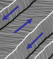
A Lorentz TEM image of domain structure in a permalloy film. Arrows indicate the field direction in the individual domains. John Cumings, University of Maryland MRSEC
Senior Investigators
- John Cumings (leader), Materials Science & Engineering
- Oded Rabin, Materials Science & Engineering
- H. Dennis Drew, Physics
- Sang-Wook Cheong, Physics, Rutgers
Microscopic imaging of magnetism is important for understanding how atomic-scale structure determines magnetic properties. This Maryland MRSEC seed project aims to develop novel magnetic imaging techniques using the transmission electron microscope (TEM), which can then be used to simultaneously measure atomic-scale structure and magnetic properties. One technique employed by the Maryland MRSEC seed team is Lorentz microscopy (see Figure), which images the local magnetic field through its effect on the TEM electron beam. The Maryland MRSEC team is applying Lorentz microscopy to in-situ imaging of the switching of magnetic structures with electric field applied to a piezoelectric. The seed team is also developing a completely new magnetic imaging technique called electron magnetic linear dichroism, or EMLD. EMLD is based on electron energy loss spectroscopy (EELS) of the near-edge structure of transition metals. The spectrosocpic signature of the 2p-3d transition is sensitive to the relative orientation of the magnetic moment and the incident electron beam. The difference signal gives the dichroism.
The seed team is working to demonstrate EMLD by imaging the perpendicular core of magnetic vortex structures. It is known that the core of these vortices forces the magnetic moment to point out-of-plane, a geometry inaccessible to Lorentz microscopy. This provides an ideal nanoscale strcuture for testing the technique, as there are few other techniques for imaging the spin in these structures. In addition to allowing the development of a new imaging technique, there is much to learn about the structure of these cores and how it depends on materials.
Highlights
- Direct Observation of Electric-Field Induced Motion of a Magnetic Domain Wall
- For the complete list, see the Highlights page
Publications
Contact Us | Page Last Updated: 05/01/09 | Site Map


